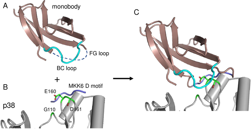
Figure 2.
Overview of anchor-guided engineering (A) Structure of a monobody (1fna) with the FG loop removed to allow insertion of the anchor peptide and the BC loop colored in cyan. (B) Crystal structure of p38α (gray) bound to the MKK6 D motif (PDB ID: 2y8o). (C) Model generated with the AnchorDesign protocol in Rosetta showing that insertion of the D motif in the FG loop of the monobody is predicted to place the BC loop adjacent to a patch of residues (green) that differ between p38α and ERK2.

