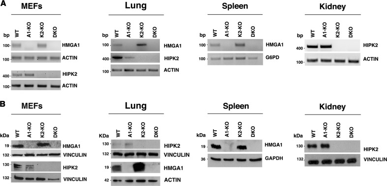
Fig. 1. Lack of HMGA1 and HIPK2 expression in A1/K2-KO mice.
a RT-PCR expression analysis of the Hmga1 and Hipk2 genes in mouse embryo fibroblasts (MEFs) at passage 3 and in lung from wild-type (WT), A1-KO, K2-KO, and DKO mice, of Hmga1 gene in spleen, and Hipk2 gene in kidney. Actin and G6pd gene expression was used as control. b Western blot analysis of HMGA1 and HIPK2 proteins in total cellular extracts from MEFs at passage 3, lung, spleen and kidney of WT, A1-KO, K2-KO, and DKO mice were performed with the indicated antibodies. Anti-actin, anti-vinculin, and anti-GAPDH were used as loading control

