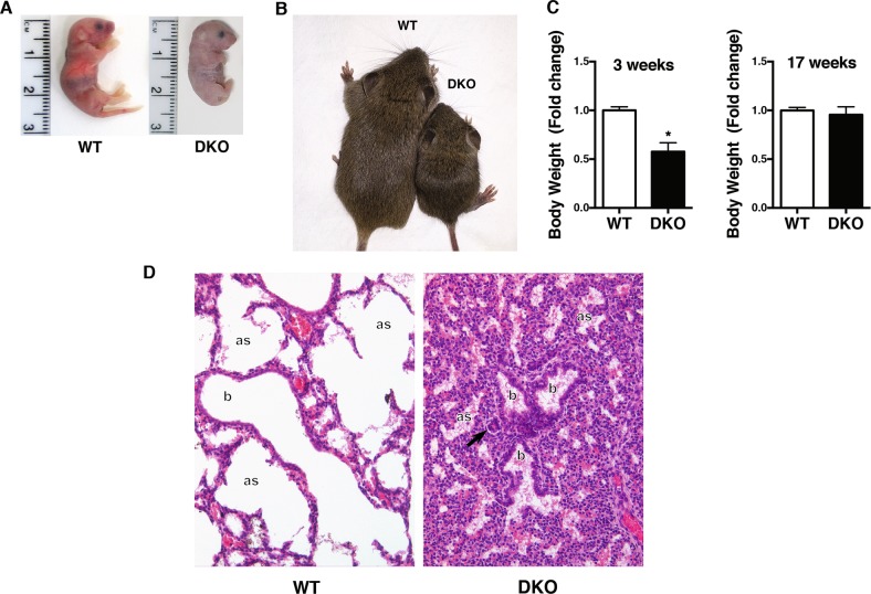
Fig. 2. DKO mice display neonatal atelectasis.
a A photograph of WT (left panel) and DKO (right panel) pups at birth. b A photograph of WT (on the left) and DKO (on the right) pups at 20 days of life. c Body weight variation of n = 8 WT (four male and four female) and n = 7 DKO (four male and three female) mice at 3 and 17 weeks of age. For statistical analysis, Student’s t test was used for each genotype. Data represent the mean ± SD * <0.05. d Representative hematoxylin and eosin staining of WT and DKO lungs of mice at P1 (×:200 magnification). The observed atelectasis was often associated with prominent vasoconstriction of peribronchiolar arterioles (arrow); as: alveolar spaces; b: terminal bronchioles

