Fig. 2.
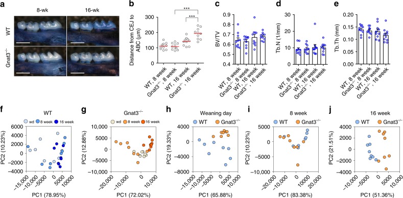
Accelerated naturally occurring alveolar bone loss and distinct commensal oral microbiota in Gnat3−/− mice. a Defleshed maxillae stained with methylene blue from wild-type (WT) and Gnat3−/− mice at 8 or 16 weeks of age. Yellow dotted line indicates the area between the cementoenamel junction (CEJ) of the second maxillary molar and the alveolar bone crest (ABC). Scale bars: 500 μm. b Quantitation of the distance from the CEJ of the second maxillary molar to the ABC. The result for each mouse is plotted; the red line indicates the mean (n = 10 mice). ***p < 0.001, one-way ANOVA test followed by Tukey’s test. c–e MicroCT analysis of alveolar bone (n = 10 mice). BV/TV bone volume/tissue volume, Tb.N trabecular number, Tb.Th trabecular thickness. f–j Principal component analysis (PCA) of microbiota recovered from oral swabs collected from WT and Gnat3−/− mice at three time points: weaning day (wd) and 8 and 16 weeks of age. Each circle represents an individual oral swab sample (n = 8 mice), color coded by age (f, g) or genotype (h–j). Error bars in c–e represent the SEM. Source data are provided as a Source Data file

