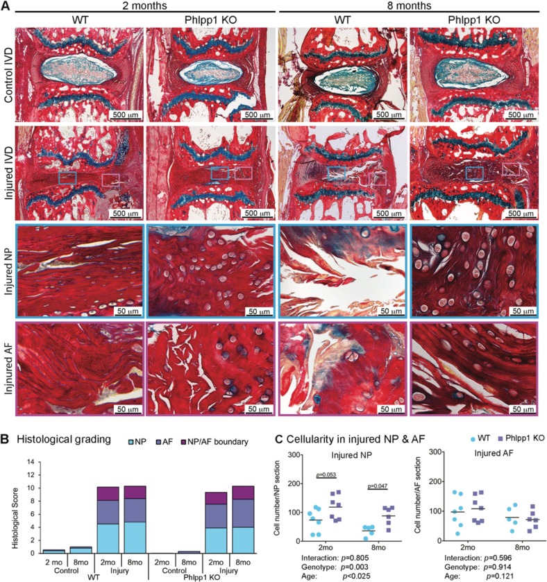
Fig. 6. Phlpp1-deletion improved NP cellularity after needle puncture injury.
a) Representative images of Picrosirius Red- Alcian Blue staining of IVD sections 2 and 8 months after injury. Green boxes indicate ROI of the NP and pink boxes indicate the AF. b) Distribution of IVD score of the NP, AF, and NP/AF boundary. IVD score was significantly increased both in WT and Phlpp1 KO at 2 (n = 7 for control WT, n = 6 for injured WT mice; n = 10 for control Phlpp1 KO and n = 8 for injured Phlpp1 KO mice) and 8 months (n = 5 per group) after injury, but no differences were observed between injured WT and Phlpp1 KO IVDs at both timepoints. c) Cellularity in NP and AF tissues after injury. The injured NP of Phlpp1 KO mice tended to be more cellular than the cellularity in WT mice both 2 (n = 7 per group) and 8 (n = 5 for WT; n = 6 for Phlpp1 KO) months after injury, and the cellularity of injured AF was similar between WT and Phlpp1 KO mice. Two-way ANOVAs were used to assess the effect of injury and genotype

