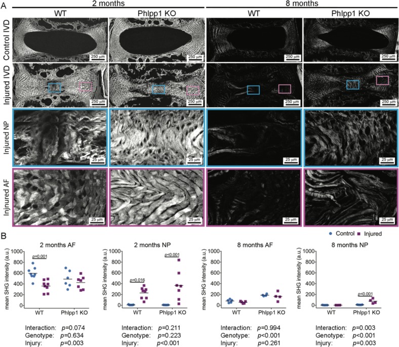
Fig. 7. Phlpp1-deletion improved collagen structure in the NP following needle puncture injury.
a) Representative images of SHG microscopy. Boxes indicate the ROI in injured levels of the NP (blue boxes) and the AF (pink boxes). b Quantification of mean SHG intensity (n = 7 per group at 2 months; n = 5 for WT and n = 4 for Phlpp1 KO at 8 months). Mean SHG intensity was reduced in WT 2 months after needle puncture, but not in Phlpp1 KO mice, whereas in the NP, it was increased in both WT and Phlpp1 KO mice. By 8 months, the general mean SHG intensity was decreased in all groups relative to the 2 months timepoint. The AF in Phlpp1 KO mice showed higher SHG intensity than the AF in WT mice and retained the tendency after injury. In the NP, SHG intensity was only increased in injured Phlpp1 KO mice, compared with all other three groups. Two-way ANOVAs were used to assess the effect of injury and genotype

