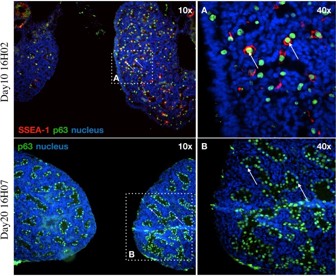
Figure 6.
Examination the gonads of the hybrid embryos: p63 and SSEA-1 immunostaining identify the endogenous PGCs in the gonads of hybrid embryo at day-10 (16H02) and p63 at day-20 (16H07). (A) The SSEA-1 expressing PGCs are red colored on the cell surface. The p63 expressing PGCs are green colored in nucleus. White square shows the cells on the picture (A) (right top). (B) The p63 expressing PGCs are green colored in nucleus. White square shows the cells on the picture (B) (right bottom). White arrows demonstrate two host derived PGCs. For nuclear staining (nucleus) we used Hoechst 33342 staining (blue).

