Fig. 2.
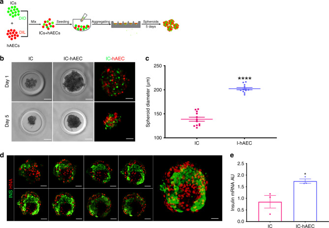
In vitro characterization of islet organoids. a Schematic representation of islet organoid engineering. After labeling with Dio and DiI, ICs and hAECs were mixed, seeded and incubated several days on 3D agarose-patterned microwells to generate islet organoids. b Phase-contrast and corresponding fluorescence views of spheroids in one microwell on days 1 and 5. After 5 day culture (bottom), cells undergo compaction and spheroids appear to acquire a smooth border as compared with aggregated cells at day 1. Scale bar = 50 μm. c Diameters of IC spheroids and IC-hAEC organoids (n = 12). ****p > 0.0001, unpaired Student’s t test. d Confocal views of islet-cell construct. Cell arrangement and composition of the islet organoid on day 14. Islet-derived cells stained for Insulin (green) and hAECs for human nuclear factor (red). Every ninth section of a z-stacked and the entire 3-D reconstructed islet heterospheroid (right panel) are shown. Scale bar = 50 μm. e Insulin mRNA expressed by IC spheroids and IC + hAEC organoids; insulin mRNA was analyzed by qPCR, arbitrary units (AU) after normalization to housekeeping genes. *p < 0.04, unpaired Student’s t test, n = 3. All data shown are mean ± SEM

