Figure 1.
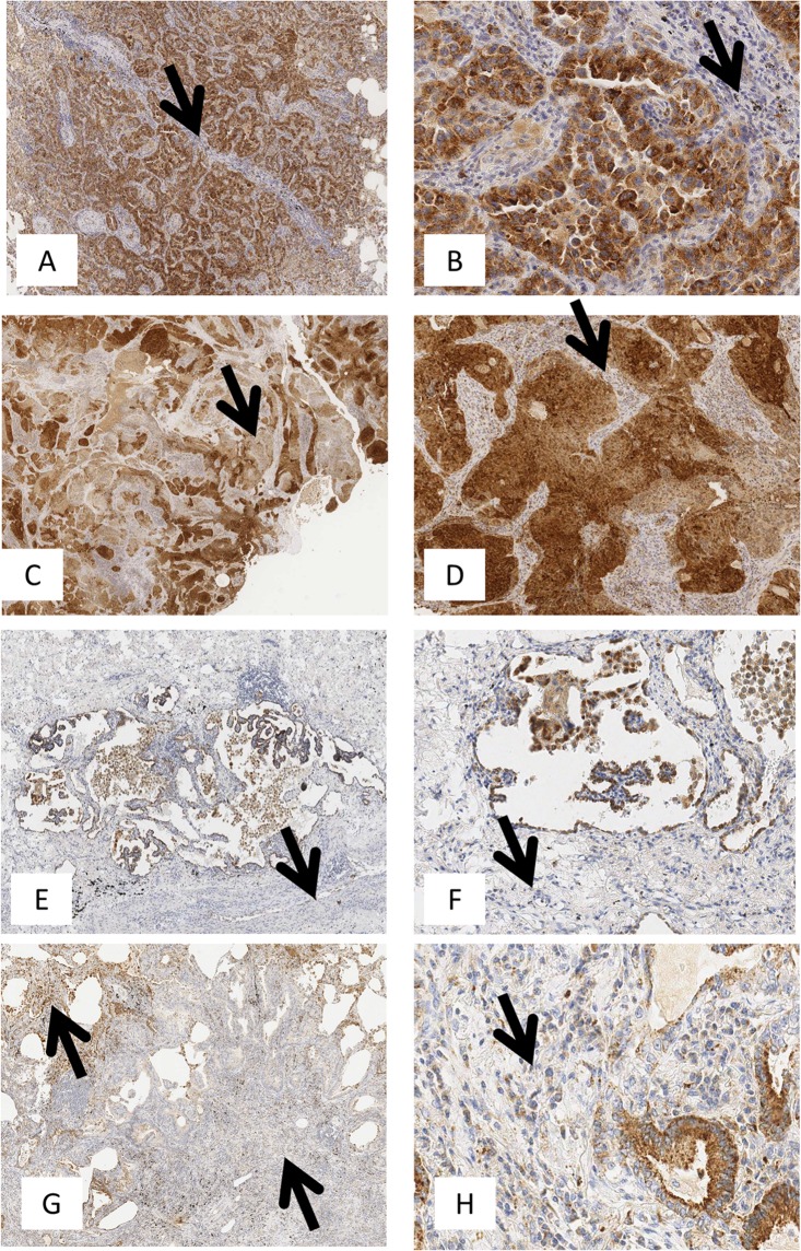
TLR3 immunohistochemical expression in lung adenocarcinoma. TLR3 immunohistochemical staining was performed on FFPE NSCLC tissue as described in Materials and Methods. TLR3 expression was defined in tumor cells and immune cells infiltrating the stroma and in tumor using a semiquantitative method that defined the percentage of positive cells of the total number of cancer or immune cells in the sample (0 = no positive cells; 1 ≤ 25%; 25% < 2 ≤ 50%; 50% < 3 ≤ 75%; 4 > 75%). (A–G) are images acquired at 100X magnification. (A) TLR3 positivity in 95% of tumor cells of the total number of cancer cells (score = 4); (B) TLR3 positivity in 65% of tumor cells of the total number of cancer cells (score = 3); (C) TLR3 positivity is 30% (score = 2) and 5% of tumor cells of the total number of cancer cells (score = 1); (D) Rare TLR3 positivity in neoplastic cells. Neoplastic cells show intense cytoplasmic staining at 200X magnification (B–F). TLR3 is abundant in neoplastic cells but is expressed in fewer than 25% of non-neoplastic stromal immune cells (B,D,F,H arrows; 200X magnification).

