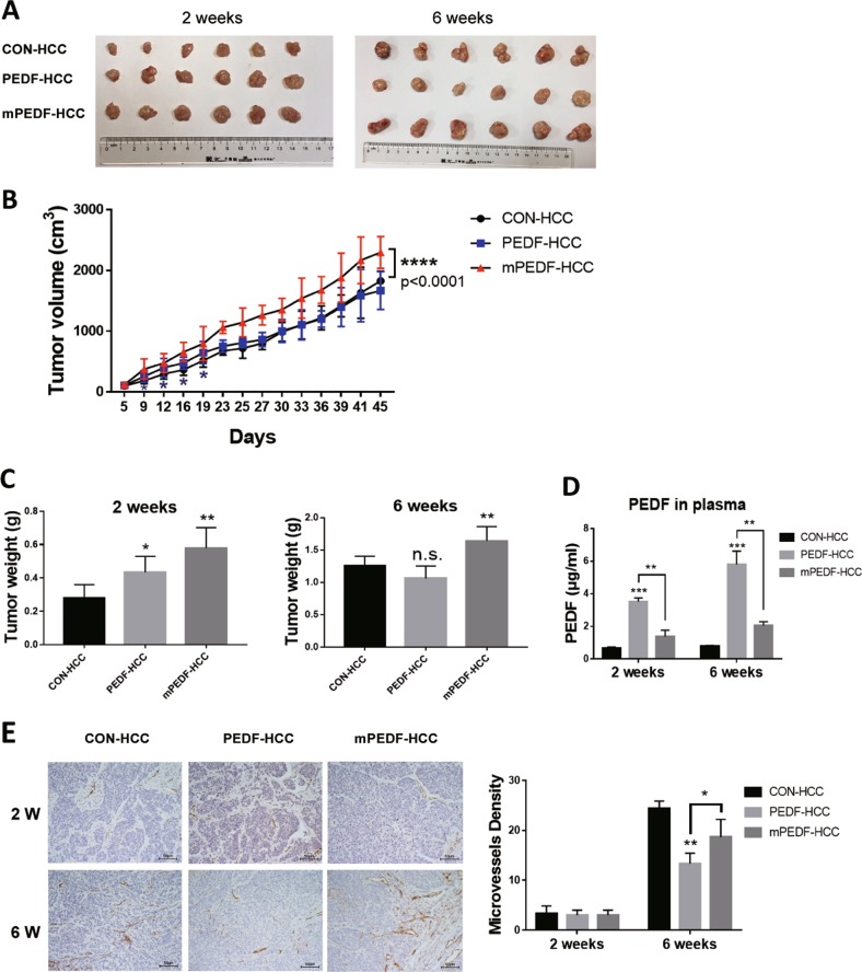
Fig. 2. Dual effects of PEDF on different stages of tumor development in HCC xenograft models.
Heterotopic HCC xenograft mouse models were performed using CON-, PEDF-, and mPEDF-HepG2 cells. Tumors were collected in the second and sixth week (n = 6). Representative images of tumors (a), the tumor growth curve (b), tumor weight (c), and secreted PEDF levels in the plasma (d) of CON-, PEDF-, and mPEDF-HCC groups at different time points were shown. e Immunostaining for CD31 and MVD assay were applied on indicated tumor xenografts collected in the second (2 W) and sixth week (6 W) (n = 3). All data are presented as mean ± SD and *p < 0.05, **p < 0.01, ***p < 0.001, and ****p < 0.0001, respectively

