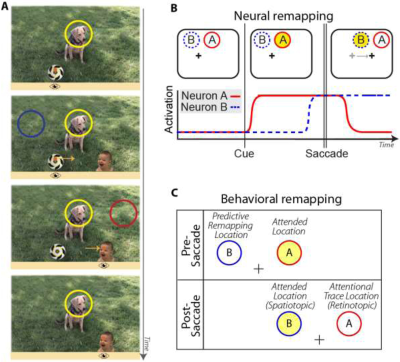
Figure 1. Dual-spotlight model of attentional remapping.
A: Real-world example illustrating sustained covert attention at a spatiotopic task-relevant location (dog), while saccading from soccer ball to baby. Orange dot indicates current fixation location (horizontal position also shown with cartoon eye icon below image); light orange arrows indicate planned or recently completed saccade trajectory. When viewer is initially fixating on soccer ball but attending to dog, the attended location (yellow circle) is in the upper right visual field. Saccading to the baby moves the dog into the upper left visual field. The dog’s spatiotopic position remains stable, but its retinotopic position has changed. From top to bottom, panels indicate early pre-saccade, later pre-saccade (predictive remapping period), early post-saccade (retinotopic trace period), later post-saccade. Red and blue circles correspond to other locations that may be attended during remapping, as defined in B,C. B: Hypothetical responses of two visual neurons with different spatial receptive fields. Yellow circle represents to-be-attended spatiotopic location. Before the saccade, the attended location falls within Neuron A’s receptive field; after the saccade, it falls in Neuron B’s. “Predictive remapping” is when Neuron B begins to respond in anticipation of the saccade. “Retinotopic attentional trace” is when Neuron A continues to respond for a period of time after the eye movement. Thus there is a period of time where both spatiotopic and retinotopic locations are facilitated. C: Corresponding locations for a behavioral study.

