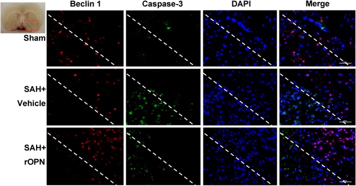
Figure 6.

rOPN administration influenced the interaction and balance between Beclin 1 and Caspase‐3 at 24 h after SAH. Double immunofluorescence staining of Caspase‐3 and Beclin 1 in Sham group, SAH + Vehicle group and SAH + rOPN group at 24 h after SAH induction. Sample size is 9, n = 3 per group. Localization of Caspase‐3 can be cytoplasmic and nuclear. Staining in the nucleus is considered to be an indication of active Caspase‐3. The dashed lines and the red box on brain slice images indicate the locations observed. Vehicle, phosphate‐buffered saline; Scale bar = 50 μm
