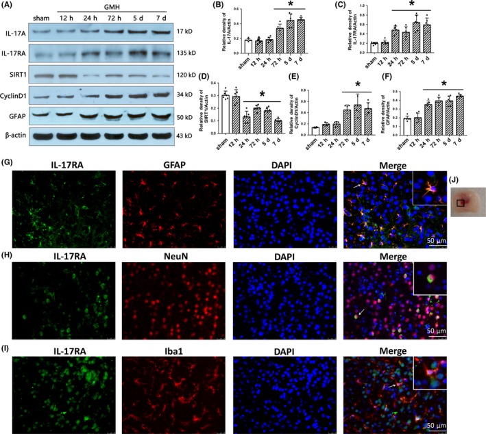
Figure 2.

The time course of IL‐17A, IL‐17RA, SIRT1, CyclinD1, GFAP, and cellular localization of IL‐17RA after GMH. A‐F, The expression of IL‐17A, IL‐17RA, SIRT1, CyclinD1, and GFAP at different times after GMH. * P < 0.05 vs sham, mean ± SD, one‐way ANOVA, Tukey's test, n = 6/group. G‐I, Colocalization of IL‐17RA with GFAP, NeuN, and Iba1 at 72 h after GMH. J, indicates the photographed location. Scale bar = 50 µm, n = 2/group. GMH, germinal matrix hemorrhage. GFAP, glial fibrillary acidic protein; NeuN, hexaribonucleotide binding protein 3; Iba‐1, ionized calcium binding adapter molecule 1; DAPI, 4′,6‐diamidino‐2‐phenylindole; IL‐17RA, IL‐17A receptor
