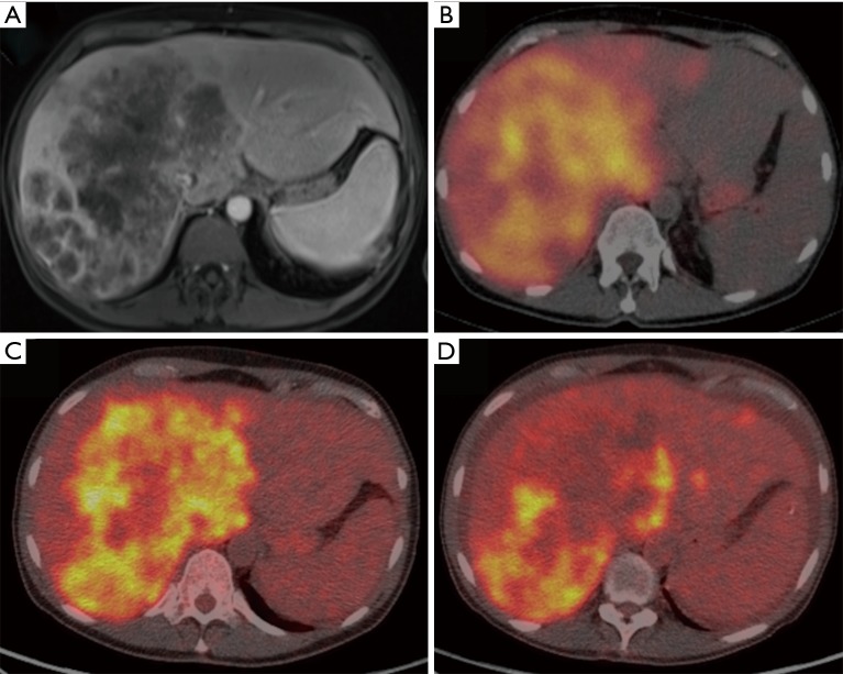
Figure 5.
Partial response by PET/CT. (A) Post-gadolinium T1-weighted MRI shows diffuse involvement of the right hepatic. (B) Diffuse Y90 deposition is shown on post-90Y-RE Bremsstrah lung SPECT/CT scan post radioembolization. (C,D) Pre- and post-90Y-RE PET scan shows SUV changes in tumor burden after 3 months from treatment. PET, positron emission tomography; CT, computed tomography; SPECT, single-photon emission computed tomography; SUV, standardized uptake value.

