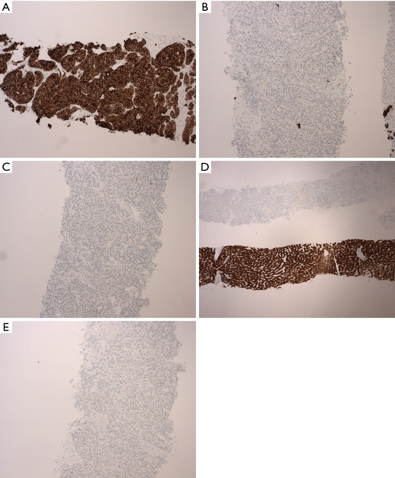
Figure 3.
Further histology slides showing diagnostic immunohistochemical stains. (A) High molecular weight (HMW) keratin staining positive in tumor cells (magnification ×40); (B) CK 7 negative staining in tumor cells (magnification ×100); (C) CK 20 negative staining in tumor cells and normal liver cells (magnification ×100); (D) arginase demonstrating positive staining in normal liver cells compared to negative staining in tumor cells (magnification ×25); (E) CD56 negative staining in tumor cells (magnification ×100). CK, cytokeratin.

