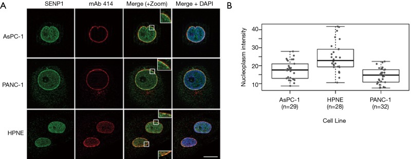
Figure 2.
SENP1 localizes to the nucleoplasm and nuclear pore complexes (NPCs). (A) Representative immunofluorescence microscopy images of pancreas-derived cells co-stained with antibodies recognizing SENP1 and NPCs (mAb 414). Scale bar is 10 µm; (B) quantitation of SENP1 nucleoplasmic signal from immunofluorescence images.

