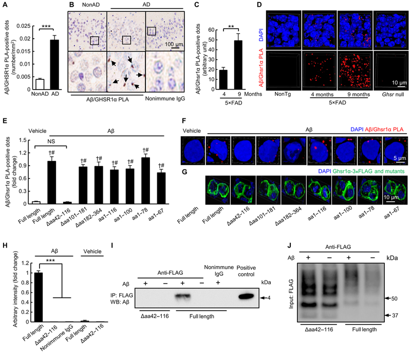
Fig. 1. Aβ physically interacts with GHSR1α.
(A) Measurement of PLA-positive dots for Aβ/GHSR1α complex in hippocampi from subjects with AD. ***P < 0.001, unpaired Student’s t test. n = 4 healthy donors or subjects with AD. (B) Representative images of quantification in (A). Arrows indicate Aβ/GHSR1α PLA-positive dots. (C) Analysis of Aβ/Ghsr1α PLA-positive dots in the hippocampal region from 4- and 9-month-old 5×FAD mice. **P < 0.01, unpaired Student’s t test. n = 4 mice per group. (D) Representative three-dimensional (3D) reconstructed images. The slices from 9-month-old Ghsr null mice were used as negative control. (E to G) Analysis of Ghsr1α/Aβ PLA-positive dots in HEK 293T cells expressing different forms of Ghsr1α treated with vehicle or 5 μM oligomeric Aβ42 for 24 hours. Anti-FLAG antibody was used to detect Ghsr1α and its mutants. †P < 0.001 versus cells expressing full-length Ghsr1α without oligomeric Aβ42 treatment and #P < 0.001 versus cells expressing Ghsr1α Δaa42–116 with oligomeric Aβ42 treatment, unpaired Student’s t test. n = 4 to 7. (F) Representative 3D reconstructed images of Ghsr1α/Aβ PLA-positive dots in HEK 293T cells expressing different forms of Ghsr1α treated with vehicle or oligomeric Aβ42 (top panels) and (G) representative 3D reconstructed images of immunofluorescent staining of different forms of Ghsr1α (bottom panels) recognized by anti-FLAG antibody. (H) Densitometry of all immunoreactive bands generated from Co-IP on HEK 293T cells expressing different forms of Ghsr1α treated with vehicle or 5 μM oligomeric Aβ42 for 24 hours. ***P < 0.001, one-way ANOVA followed by Bonferroni post hoc analysis. Data were collected from three independent experiments. n (from left to right) = 3, 5, 2, 3, and 3. Nonimmune immunoglobulin G (IgG) to replace specific FLAG antibody was used for examining specificity of Co-IP. (I) Representative immunoblots showing the interaction of oligomeric Aβ42 with Ghsr1α and Ghsr1α Δaa42–116. (J) Representative immunoblots showing the input of Ghsr1α and Ghsr1α Δaa42–116. DAPI, 4′,6-diamidino-2-phenylindole; NS, not significant; IP, immunoprecipitation; WB, Western blot.

