Figure 1.
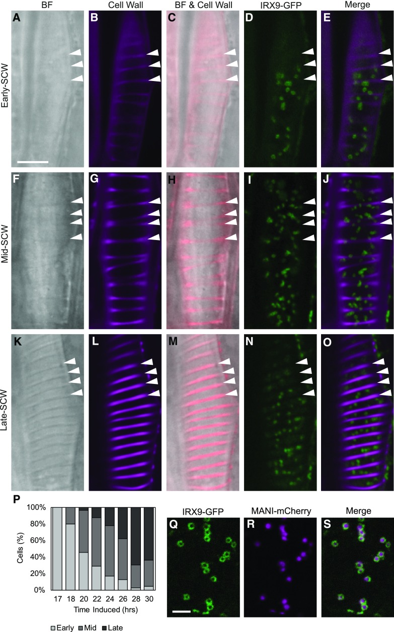
IRX9-GFP localizes to the Golgi throughout SCW deposition. A to O, IRX9-GFP and SCW localization at the early-SCW, mid-SCW, and late-SCW stages. SCW bands are visible in brightfield (BF) in mid-SCW and late-SCW stages, and are sometimes also apparent when early-SCW cells are stained with propidium iodide (Cell Wall). IRX9-GFP is found in Golgi-like punctae in all stages. Cell wall images were acquired using different sensitivities. Images are representative single optical sections through the cell cortex. Arrowheads = SCW bands. Scale bar = 10 µm. P, The stage of SCW development varies among populations of transdifferentiating cells. The percent of IRX9-GFP expressing cells assigned to the early, mid, or late stages of SCW-deposition at each time point following treatment with DEX. Included per induction time point were 60–204 cells, across 4 replicate experiments. Q to S, IRX9-GFP and MANI-mCherry colocalize in the Golgi apparatus. Spinning disk confocal colocalization of the xylan biosynthetic IRX9-GFP (Q) and the Golgi-marker MANI-mCherry (R). The co-occurrence of the proteins in every Golgi stack is apparent in the merged image (S). Distinct distributions within the Golgi are visible as ring-shaped IRX9-GFP and solid MANI-mCherry signals. Scale bar = 5 µm.

