
Figure EV3. CENP‐A nucleosome/CENP‐CCR complex structure.
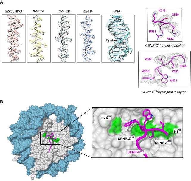
- Representative cryo‐EM densities showing fitted model for DNA and each of the histones (left), arginine anchor, and hydrophobic regions of CENP‐CCR (right).
- Surface representation of nucleosome (histone core—gray, DNA—blue), showing a hydrophobic groove (green) on the nucleosome formed by H2AL108, CENP‐AL135, CENP‐AL139, and H4V60. CENP‐CCR is shown as a purple coil with hydrophobic sidechains in stick representation.