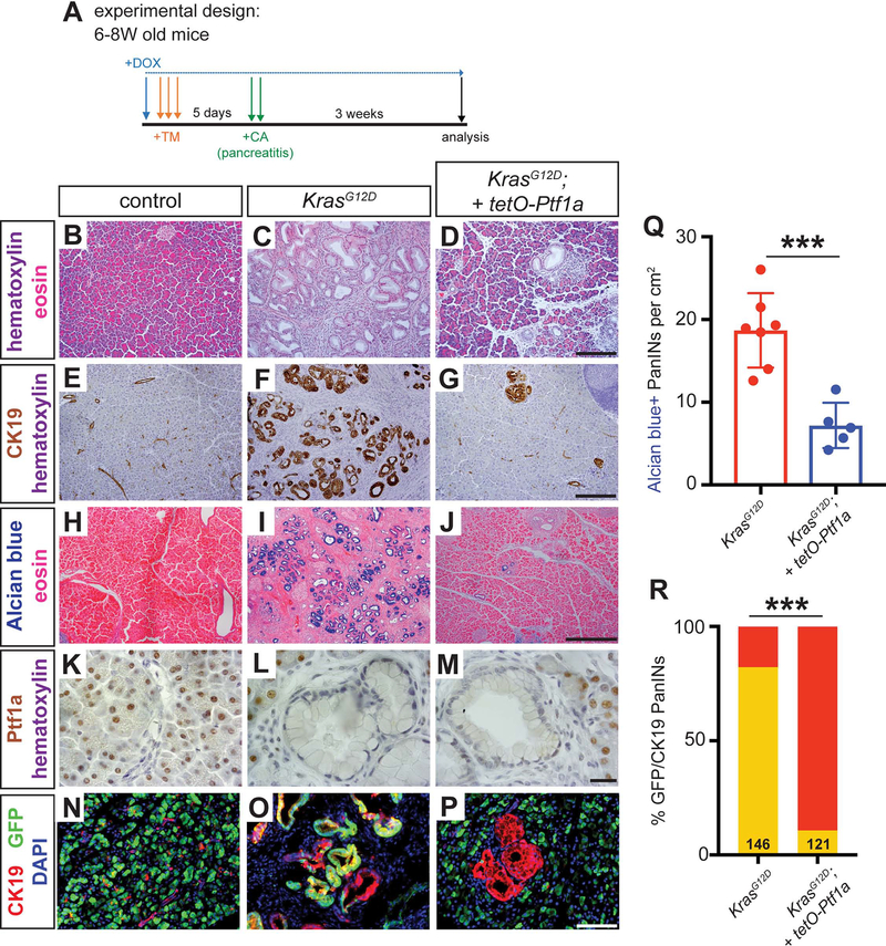
Figure 3. Maintenance of acinar identity inhibits inflammation-induced PanIN formation.
(A) Mice of indicated genotypes were started on DOX (1 mg/ml in drinking water) 24 hours before TM administration (0.25 mg/g body weight, daily over 3 consecutive days). Five days following the final TM dose, mice were administered six hourly injections of 0.1 μg/g caerulein on two consecutive days. Mice were euthanized for pancreatic histology at three weeks following the final caerulein injection. (B-D) H&E staining of pancreata from mice of indicated genotypes 3 weeks after caerulein-induced pancreatitis (20x, scale bar = 200 μm). (E-G) IHC for CK19 (20x, scale bar = 200 μm) and (H-J) Alcian blue staining (10x, scale bar = 500 μm), highlighting PanIN formation. (K-M) IHC for Ptf1a (100x, scale bar = 25 μm), showing lack of nuclear Ptf1a in normal ducts of control mice as well as in PanINs, compared to adjacent acinar cells. (N-P) Immunofluorescence for the duct marker CK19 (red), GFP (R26rtTA. green) and DAPI (blue), highlighting CK19+/GFP-negative escaper PanIN cells in KrasG12D; tetO-Ptf1a pancreata (20x, scale bar is 100 μm). (Q) Quantification of the genotype-dependent PanIN burden, indicating significant reduction in KrasG12D; tetO-Ptf1a pancreata (unpaired t-test, P<0.01). (R) Proportion of CK19+/GFP+ dual-positive PanINs (Fisher’s exact test, P<0.001).

