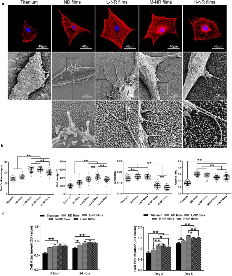
Fig. 2.
Cellular adhesion behavior on various substrates. a Upper, immunofluorescence observation of cellular cytoskeletonal morphology by staining the actin (red), and nuclei (blue). Middle, SEM observation of the typical cellular pseudopodia. Lower, the corresponding local amplification of filopodia (white arrows). b The corresponding quantitative analysis of cellular Feret’s dimeter, average cell area, circularity and aspect ratio. c Cellular viability analysis (CCK-8 assay) for cellular adhesion (6 and 24 h) and proliferation (2 and 5 days)

