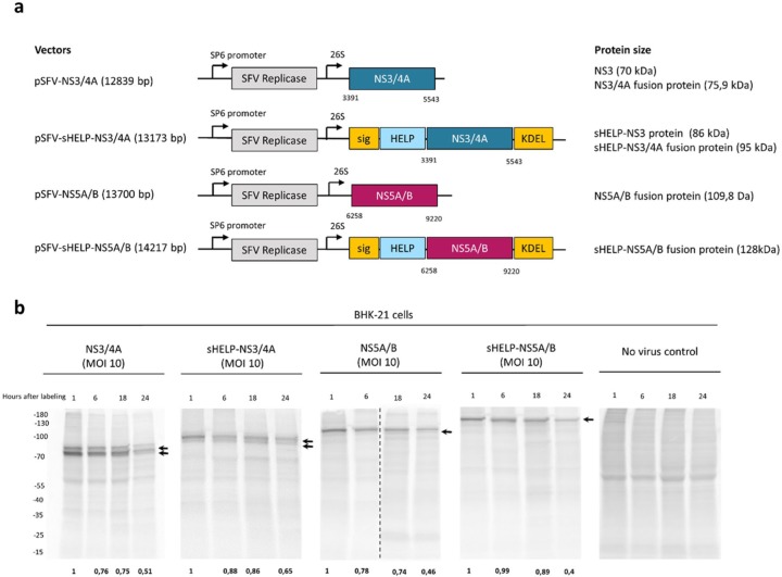
Figure 1.
Expression and stability of HCV NS3/4A and NS5A/B proteins in vitro. (a) Schematic representation of the plasmid SFV DNA vectors and the corresponding antigens (NS3/4A or NS5A/B). The numbers indicate the nucleotide position in the plasmid DNA containing the full genome of HCV 1a (H/FL). SP6 promoter; 26S, subgenomic 26S promoter. (b) BHK-21 cells were incubated with SFV replicon particles encoding for NS3/4A, sHELP-NS3/4A, NS5A/B, sHELP-NS5A/B or without SFV at an MOI of 10. Cells were pulsed with [35S]-methionine/cysteine for 1 h after incubation with the SFV particles for 6 h. Cell lysates were collected at 1, 6, 18, and 24 h post [35S]-methionine/cysteine pulse-chase labeling. Radioactively labeled proteins were revealed by autoradiography after 12% SDS-PAGE. Vertical dotted lines separate different gels that run at the same time. Arrows indicate the expression of the corresponding transgene-encoded proteins. Numbers at the bottom of the gels show remaining expression of the total transgene-encoded proteins when compared with their expression at time point 1 h after labeling. Data represent results from two independent experiments.

