
Fig. 8.
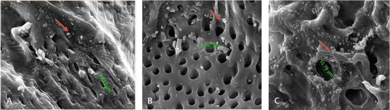
Scanning electron microcopy micrographs showed adipose-derived mesenchymal stem cells seeding with bovine teeth scaffold at various time intervals: ( A ) 1, ( B ) 12, and ( C ) 24 hours of cells seeding DDM scaffold (×5,000 magnification). The arrow-marked region is the attachment point.