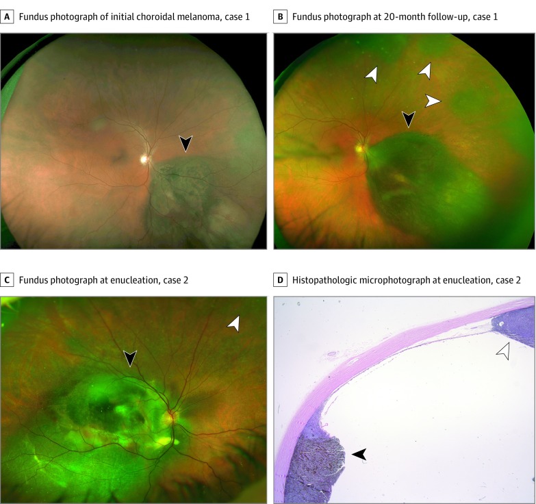
Figure 1. Patients 1 and 2 With Multifocal Uveal Melanoma.
Fundus photographs of case 1 at presentation of the initial choroidal melanoma (black arrowhead) (A and B) and at the detection of 3 distinct choroidal melanocytic tumors (white arrowheads) 20 months after plaque radiotherapy (B). Fundus photograph (C) and histopathologic photomicrograph (D) of case 2 at the time of enucleation, illustrating the original tumor treated by plaque radiotherapy (black arrowheads), and the subsequent noncontiguous tumor (white arrowheads).

