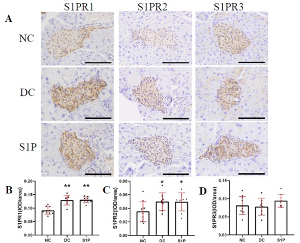
Figure 5.

Immunohistochemical staining of S1PR1-3 subtypes in the mouse islets. (A) Representative images of immunohistochemical staining of S1PR1, S1PR2 and S1PR3 in the NC (n=10), DC (n=8), and S1P (n=9) groups. S1PR1-3 expression is indicated by the brown staining in the cells. Magnification, ×400; scale bar, 100 µm. IOD/area of (B) S1PR1, (C) S1PR2 and (D) S1PR3 in all groups. Values are expressed as the mean ± standard deviation. Parametric one-way analysis of variance followed by Least Significant Difference post hoc test was performed for the comparison of the groups. *P<0.05 and **P<0.01 vs. NC. NC, normal control; DC, diabetic control; S1P, sphingosine-1-phosphate; S1PR, S1P receptor; IOD, integrated optical density.
