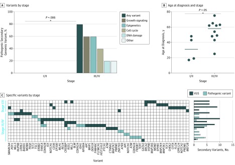
Figure 3. Prevalence of Pathogenic Secondary Genomic Variants.
A, Stage III/IV tumors were more likely to harbor a pathogenic secondary genomic variant. B, Stage III/IV tumors were associated with older age at diagnosis. Horizontal lines indicate group median. C, Heatmap of secondary genomic variants with total secondary variants noted on the rightmost y-axis. VUS indicates variant of unknown significance.
aThree participants clinically diagnosed with stage III/IV epithelioid hemangioendothelioma with genomic profiling data available but lacking confirmation of a WWTR1-CAMTA1 fusion were included in a secondary clinical assessment. These participants exhibited similar characteristics to other participants previously described.

