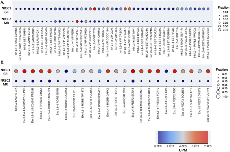
Figure 1.
Expression of GR and MR in all individual 45 inhibitory and 24 excitatory neuronal cell types of the human temporal cortex. (A) GABA-ergic (‘inh’, or inhibitory) neurons show variable expression of GR and MR. Two GABA-ergic neurons show higher expression of MR than of GR: layer 2-6 VIP/OPCT positive neurons and layer 1-2 VIP/PCDH20 positive neurons. In most cell types, receptors are expressed at intermediate to low levels. (B) Glutamatergic (‘Exc’, or excitatory) neurons generally express high levels of GR and low levels of MR. Exonic expression is shown. Color codes for counts per million (CPM) transcripts on a linear scale, and size indicates the fraction per cell types with expression >1 CPM. Data and image are from http://celltypes.brain-map.org/rnaseq/human.

