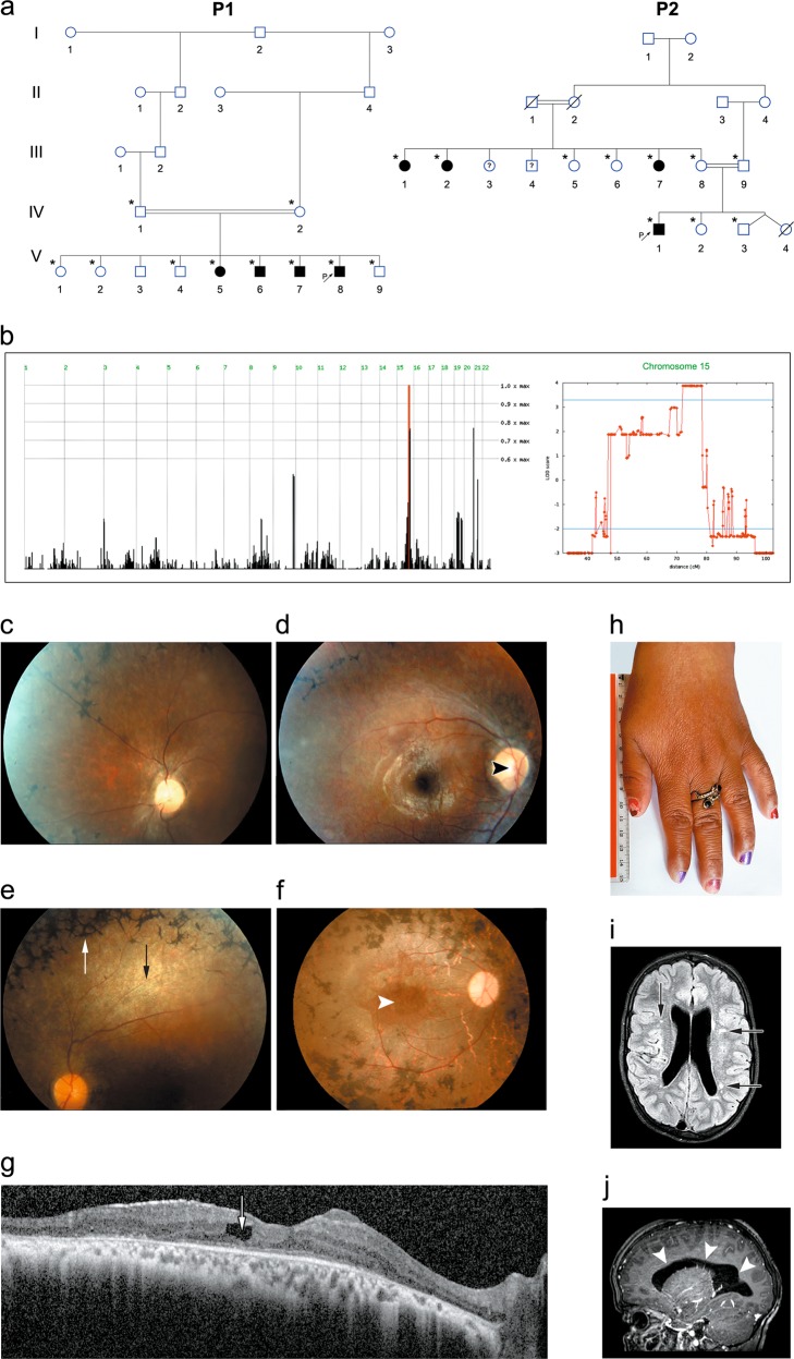
Fig. 1.
Pedigree, homozygosity mapping and disease phenotype: a Pedigrees of consanguineous Bedouin kindred studied (P1—pedigree 1, P2—pedigree 2). Subjects available to the study are marked with asterisks. All eight affected individuals were homozygous for the SCAPER (NM_020843.2) c.2806delC; p.(L936*) mutation. The mutation segregated as expected for a recessive trait. b Homozygosity-Mapper plot and LOD score analysis for chromosome 15 using SUPERLINK online. c–e Fundus photographs of three P1 patients: c P1:V7 (14 years) (left eye), d P1:V6 (27 years) (right eye), e P1:V5 (33 years) (left eye), showing diffuse chorio-retinal degeneration, as evidenced by attenuated arterioles (black arrow), granular appearance of the retina with hypopigmented spots, optic disk pallor (black arrowhead) and a typical bone-spicule type hyperpigmentation (white arrow) in the retinal mid-periphery. f Fundus photograph of the right eye of patient P2:III2 (47 years), showing optic atrophy, attenuated vessels, gray atrophic retina with diffuse coarse pigment clumps and bone spicules, as well as a severe maculopathy (white arrowhead) seen also on the SD-OCT image of this eye. g Irregular and thickened foveal margins and intra-retinal fluid with cystoid macular edema (arrow) (patient P2:III2). h Brachydactyly with tapering fingers (patient P2:III1). i, j Brain MRI of patient P2:IV1 at the age of 10 years, demonstrating mildly enlarged lateral ventricles (i; arrows) and several loci of irregular signal in the brain parenchyma (j; arrowheads) above the tentorium, in the posterior white matter and along the ependyma

