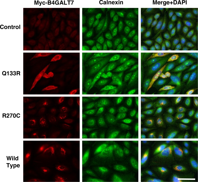
Fig. 5.
Localization of myc-(B4GALT7) variants with calnexin, a marker for the endoplasmic reticulum. Immunostaining with anti-calnexin (green) and anti-myc-tag antibody (red) in untransfected pgsB-618 cells and pgsB-618 cells stably transfected with B4GALT7-WT, B4GALT7-Q133R, or B4GALT7-R270C. Merge + DAPI shows a three-color panel with DAPI, a blue nuclear stain, calnexin (green), and myc-B4GALT7 (red). Bar is 31 µm for all panels

