Figure 6.
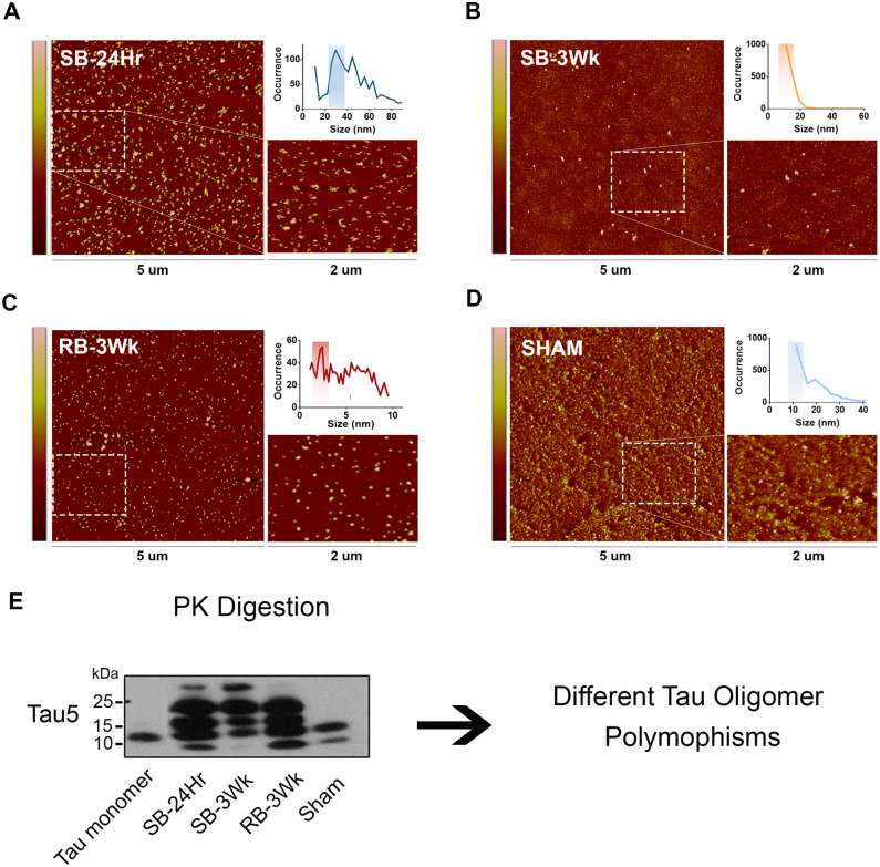
Tau oligomers derived from brains exposed to single versus repetitive TBI represent different tau polymorphisms. TBI brain-derived tau oligomers were analysed using AFM. (A) SB-24Hr, (B) SB-3Wk, (C) RB-3Wk and (D) Sham oligomers show different oligomeric morphologies and size distribution profiles (shown by the graphical inlets on the top right of each panel). (E) Western blot representing the oligomers after PK treatment, probed with Tau5, total tau antibody. The samples show different banding patterns after PK digestion indicating different tau oligomeric polymorphisms.

