Figure 2.
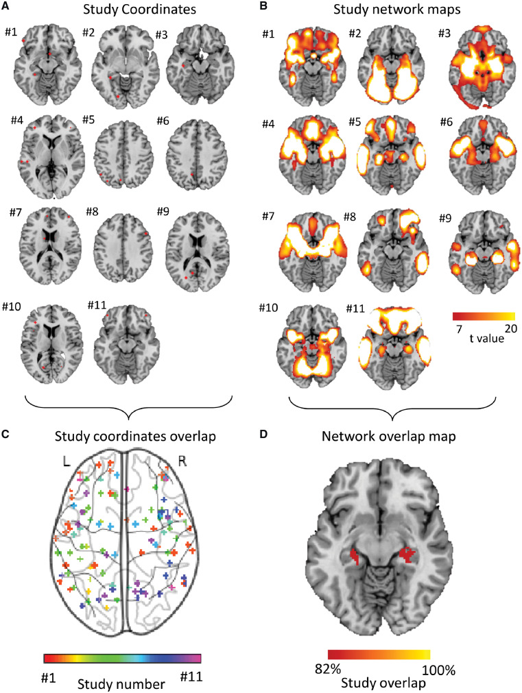
Heterogeneous neuroimaging findings in Parkinson’s disease dementia are part of a common brain network centred on the hippocampus. (A) Study coordinates. Location of coordinates for each study of Parkinson’s dementia compared with Parkinson’s without cognitive involvement. Spherical seeds were generated at each reported significant coordinate for each study of PD dementia, then added together to create one map of neuroimaging findings for each study. Numbers refer to the study number as listed in Table 1. (B) Study network maps. Regions significantly connected to each study’s neuroimaging findings were calculated using a large (n = 1000) normative connectome, creating a network map for each study (FWE-corrected P < 10−6). Locations of network connectivity for each study of Parkinson’s dementia compared with Parkinson’s without cognitive involvement. (C) Study coordinates overlap. Combined location of all coordinates across all studies of PD dementia shows pronounced heterogeneity. Each study is represented by a different colour. (D) Network overlap map. Network maps from each study were overlaid to identify functional connections common to the greatest number of studies in a whole-brain analysis. Over 80% of studies were functionally connected to the bilateral hippocampus. Section at z = −16 is shown.

