Figure 3.
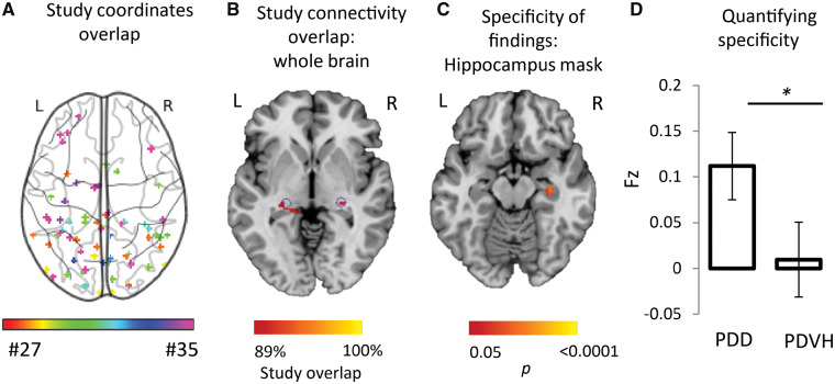
Heterogeneous neuroimaging findings in Parkinson’s disease hallucinations are part of a different brain network than PD dementia, centred on the lateral geniculate nucleus. (A) Combined location of all coordinates across all studies of Parkinson’s with visual hallucinations shows pronounced heterogeneity. Each study is represented by a different colour. (B) Connectivity maps (across the whole brain) for each study of PD hallucinations were generated and overlaid, showing network overlap in the lateral geniculate nuclei bilaterally. Section shown is at z = −4. Blue circles indicate location of lateral geniculate nucleus based on published coordinates (Burgel et al., 2006). (C) Direct comparison of network maps generated from studies of PD dementia and PD hallucinations shows specificity of hippocampal connectivity to studies of PD dementia. Map is masked to the hippocampi and FWE-corrected P < 0.05. Section shown is at z = −16. (D) Connectivity to our a priori ROI in the hippocampus was significantly stronger for studies of PD dementia compared to studies of PD hallucinations. Coordinate and network maps for all studies can be viewed in Fig. 5. * P < 0.05; PDD, Parkinson’s disease dementia; PDVH, Parkinson’s disease with visual hallucinations.

