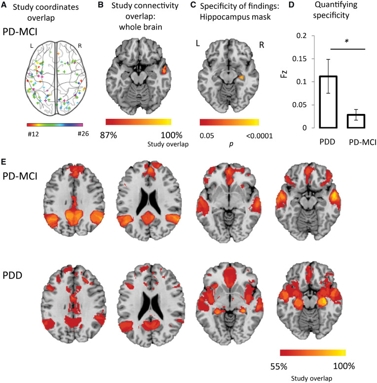
Figure 4.
Heterogeneous neuroimaging findings in Parkinson’s disease MCI are part of a network centred on posterior nodes of the default mode network. (A) Combined location of all coordinates across all studies of Parkinson’s mild cognitive impairment (PD-MCI) shows pronounced heterogeneity. Each study is represented by a different colour. (B) Connectivity maps for each study of PD-MCI were generated and overlaid, showing peak network overlap in the lateral temporal cortex. Section shown is at z = −18. (C) Direct comparison of network maps generated from studies of PD dementia and PD-MCI shows specificity of hippocampal connectivity to studies of PD dementia. Map is masked to the hippocampi and FWE-corrected P < 0.05. Section shown is at z = −14. (D) Connectivity to our a priori ROI in the hippocampus was significantly stronger for studies of PD dementia compared to studies of PD-MCI. (E) At lower network overlap thresholds, there are similarities between PD-MCI and PD dementia. This suggests that posterior nodes of the DMN are affected in both PD-MCI and PD dementia, and that at later stages, once PD dementia takes hold, hippocampal networks are affected. Sections shown are at z = 30, z = 21, z = −7 and z = −16. Coordinate and network maps for all studies can be viewed in Fig. 5. * P < 0.05; PDD, Parkinson’s disease dementia; PD-MCI, Parkinson’s disease with mild cognitive impairment.

