Figure 2.
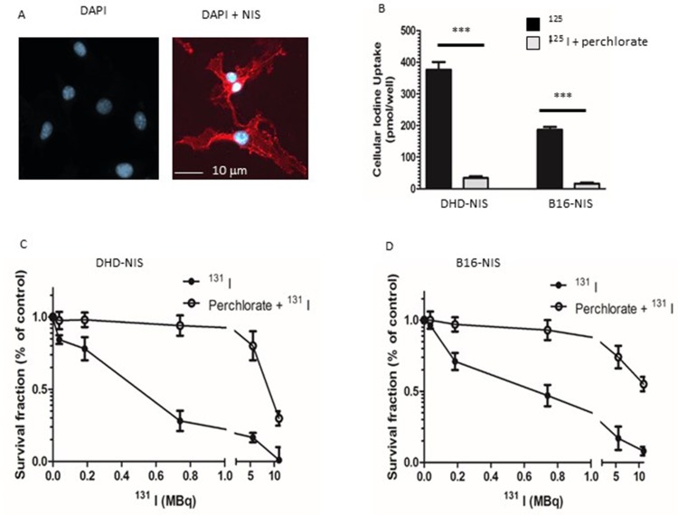
Sensitivity of NIS-expressing-B16 melanoma and -DHD colorectal carcinoma cells to radioiodine. (A) Immunofluorescence analysis of NIS expression in stably transfected B16-NIS cells. The expression of NIS at the cell surface was evaluated by using a specific anti‐mouse NIS polyclonal antibody (right panel) or rabbit IgG (left panel). (B) Cellular iodine uptake by the DHD-NIS and the B16-NIS tumor cells. DHD-NIS or B16-NIS cells were incubated for 1 hr with 125I in the presence or the absence of sodium perchlorate, a specific NIS inhibitor before being washed and lysed. Aliquots of lysates were counted in a γ counter. The data presented are the mean±standard error of the mean (SEM) of triplicates and are representative of three independent experiments. Radiation cell survival curves for DHD-NIS cells (C) and for B16-NIS cells (D). Clonogenic assays were performed on DHD-NIS or B16-NIS cells after treatment with 131I alone (0 to 11 MBq for 4 hrs) or with a combination of sodium perchlorate and 131I. ***p<0.001.

