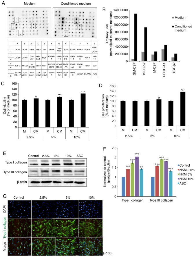
Figure 1.
Impact of NK-CdM on NHDF growth and collagen synthesis. Growth factor array analysis of NK-CdM. (A) List of growth factors on the array membrane (lower panel) with significantly detected growth factors marked (upper panel). (B) Growth factor secretion profiles of NK-CdM. A human growth factor antibody array was used to detect paracrine factors present in NK-CdM. (C) Proliferation of NHDFs treated with NK-CdM at various concentrations for 24 h. (D) Time course of NHDF proliferation up to 72 h. Data are shown as the mean ± standard deviation of three independent experiments. (E) Western blotting and (F) graphical representation of the effects of NK-CdM on collagen synthesis in NHDFs. NHDFs were treated with medium (Control), ASC, (200 μM), or NK-CdM (2.5, 5 and 10%) for 72 h and the expression of type I collagen and type III collagen was measured by western blot analysis and normalized to the expression of β-actin. (G) Immunocytochemical analysis showing the activation of type I collagen in NK-CdM-treated NHDFs. NHDFs were fixed and then incubated with a goat anti-rabbit type I collagen antibody. Type I collagen staining was detected with a fluorescein isothiocyanate conjugated anti-goat IgG antibody by fluorescence microscopy. Magnification, ×100. **P<0.01 and ***P<0.001 vs. control. NK-CdM, natural killer cell conditioned medium; NHDF, neonatal human dermal fibroblasts; ASC, ascorbic acid.

