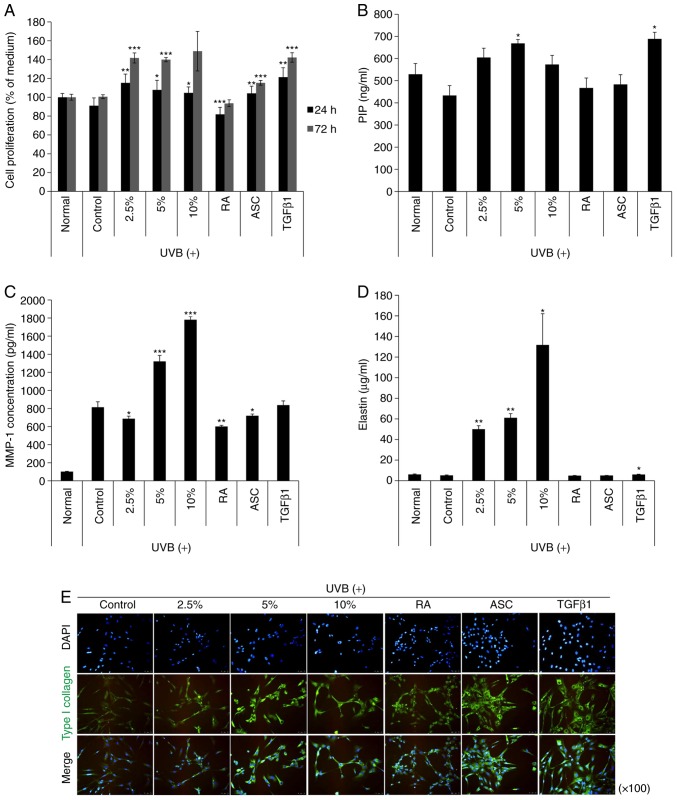
Figure 2.
The effects of NK-CdM on UV-B-induced MMP-1 and type I procollagen secretion in NHDFs. NHDFs were irradiated with 30 mJ/cm2 UV-B, followed by treatment with the indicated concentrations of NK-CdM (2.5, 5 and 10%), RA (0.03 μg/ml), ASC (200 μM), and TGF-β1 (10 ng/ml). (A) Time course of NHDF proliferation up to 24 and 72 h. (B) PIP secretion, (C) MMP-1 levels, and (D) Elastin levels in harvesting culture media at 48 h. MMP-1, PIP, and elastin levels were measured using a commercially available ELISA kit, as described in the Methods section. The data are the mean ± standard deviation values of three individual experiments. (E) Immunocytochemical analysis showing an inhibition of type 1 collagen induction in NK-CdM-treated NHDF cells. Magnification, ×100. *P<0.05, **P<0.01 and ***P<0.001 vs. Control (UV-B only group). MMP, matrix metalloproteinase; PIP, pro-collagen type 1 C-peptide; NK-CdM, natural killer cell conditioned medium; NHDF, neonatal human dermal fibroblasts; UV, ultraviolet; RA, retinoic acid; ASC, ascorbic acid; TGF, transforming growth factor.


