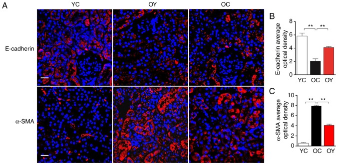
Figure 3.
Effects of incremental load training on the expression of the E-cadherin and α-SMA in the kidney tissues of aged mice. (A) Expression of E-cadherin and α-SMA were identified by immunofluorescence staining (magnification, ×400; scale bar=20 μm). The red color represents a positive signal; cell nuclei were counterstained with Hoechst (blue). Relative OD values of (B) E-cadherin and (C) α-SMA staining are shown in bar graphs. Values are expressed as the mean ± standard deviation (n=3). **P<0.01. YC, young control group; OC, elderly control group; OY, elderly exercise group; OD, optical density; α-SMA, α-smooth muscle actin.

