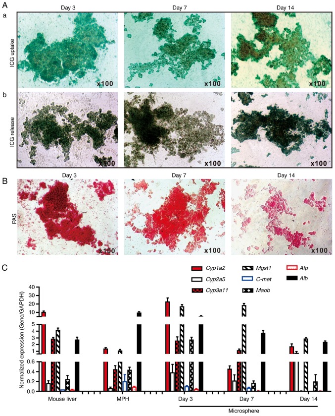
Figure 3.
Hepatic functional characterization of 3-dimensional LMTC model. (A) ICG uptake and release assay. (a) The freshly recovered mouse liver sift80 microsphere tissue was incubated with ICG (1 mg/ml in 2% BCS/DMEM) at 37°C for 30 min. ICG uptake was recorded under a bright field microscope. (b) The microsphere tissue was then incubated in 2% BCS/DMEM for an additional 3 h, and the ICG release was recorded under a bright field microscope. Each assay condition was performed in triplicate. Representative results are presented. (B) PAS staining for hepatic glycogen storage. At the indicated time points, the cultured sift80 microsphere tissue was subjected to PAS staining, and the PAS staining was recorded under a bright field microscope. Each assay condition was performed in triplicate. Representative results are presented. (C) Expression profile of hepatocyte-specific genes in freshly isolated mouse liver tissues, the MPH and the LMTC microsphere tissue cultures at different time points. Total RNA was isolated from the 6-week old C57BL mouse liver, the MPH, and microsphere tissue cultured for 3, 7 and 14 days, and subjected to touchdown quantitative polymerase chain reaction analysis to detect the expression of hepatic genes. All samples were normalized with respective GAPDH expression levels. Each assay condition was performed in triplicate. LMTC, liver microsphere tissue culture; ICG, indocyanine green; DMEM, Dulbecco's modified Eagle's medium; PAS, Periodic acid-Schiff; MPH, mouse primary hepatocytes; Cyp1a2, cytochrome P450 family 1 subfamily Amember; Cyp2a5, cytochrome P450 2A5; Cyp3a11, cytochrome P450 family 3 subfamily A member 4; Mgst1, microsomal glutathione S-transferase 1; C-Met, MET proto-oncogene, receptor tyrosine kinase; Maob, monoamine oxidase B; Afp, α fetoprotein; Alb, albumin.

