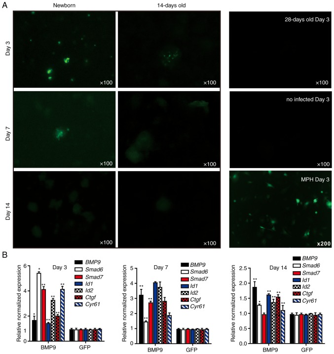
Figure 4.
3-Dimensional liver microsphere tissue culture model for hepatic exogenous investigation. (A) Cultured liver tissue was relatively refractory to Ad-mediated transgene delivery. The microsphere tissue samples were prepared from newborn, 14- and 28-day-old C57BL mouse liver samples, and were infected with the same titer of Ad-GFP. The GFP signal was recorded at 3, 7 and 14 days after infection. The MPH were also infected with Ad-GFP as a control, and the GFP signal at day 14 is presented. (B) Stimulation of the sift80 tissue microspheres with BMP9-conditioned medium to examine cell signaling and TqPCR analysis. Total RNA was isolated from the sift80 tissue microspheres treated with BMP9 or GFP conditioned medium at the indicated time points, and subjected to TqPCR analysis of BMP9 downstream target genes. All samples were normalized with respective GAPDH expression levels. *P<0.05 and **P<0.01 vs. GFP groups. Each assay condition was done in triplicate. Ad, adenovirus; GFP, green fluorescent protein; MPH, mouse primary hepatocytes; BMP9, bone morphogenic protein 9; TqPCR, touchdown quantitative polymerase chain reaction analysis; Id1, inhibitor of DNA binding 1, HLH protein; Id2, inhibitor of DNA binding 2; Ctgf, connective tissue growth factor; Cyr61, cellular communication network factor 1.

