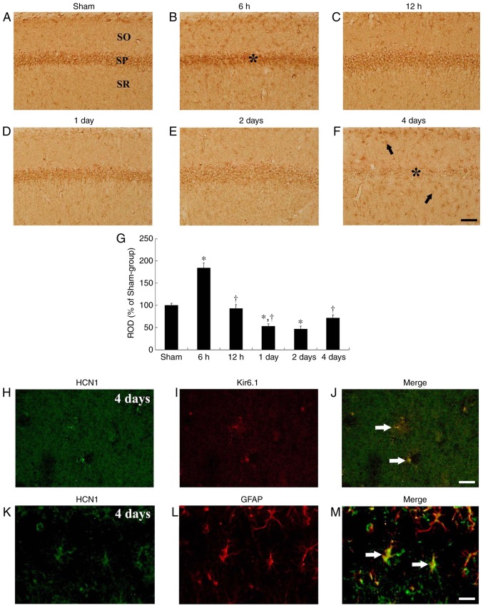
Figure 3.
HCN1 immunohistochemistry in the hippocampal CA1 region. (A-F) HCN1 immunohistochemistry in the CA1 subfield of the (A) sham and (B-F) ischemia-operated groups at (B) 6 h, (C) 12 h, (D) 1 day, (E) 2 days and (F) 4 days. HCN1 immunoreactivity was markedly increased in the CA1 pyramidal neurons 6 h after tgCI. Thereafter, HCN1 immunoreactivity is decreased in a time-dependent manner and was barely observed in the CA1 pyramidal neurons 4 days after tgCI. Scale bar, 50 µm. The asterisks and the arrows represent CA1 pyramidal neurons and non-pyramidal cells, respectively. (G) ROD is presented as percentages of HCN1 immunoreactive structures in the CA1 subfield following tgCI (n=7 at each point in time. *P<0.05 vs. the sham-operated group, †P<0.05 vs. the previous timepoint group. Bars indicate the means ± standard error of the mean. (H-M) Double immunofluorescence staining for (H and K) HCN1 (green), (I) Kir6.1 (red), (L) GFAP (red) and (J and M) merged images in the stratum pyramidale 4 days after tgCI. HCN1 immunoreactivity was localized in Kir6.1-immunoreactive pericytes and GFAP-immunoreactive astrocytes. Arrows denote Kir6.1-immunoreactive pericytes and GFAP-immunoreactive astrocytes in the respective images. Scale bar, 20 µm. HCN1, hyperpolarization-activated cyclic nucleotide-gated 1; CA1, Cornu Ammonis 1; tgCI, transient global cerebral ischemia; ROD, relative optical density; Kir6.1, ATP-sensitive inward rectifier potassium channel 8; GFAP, anti-glial fibrillary acidic protein.

