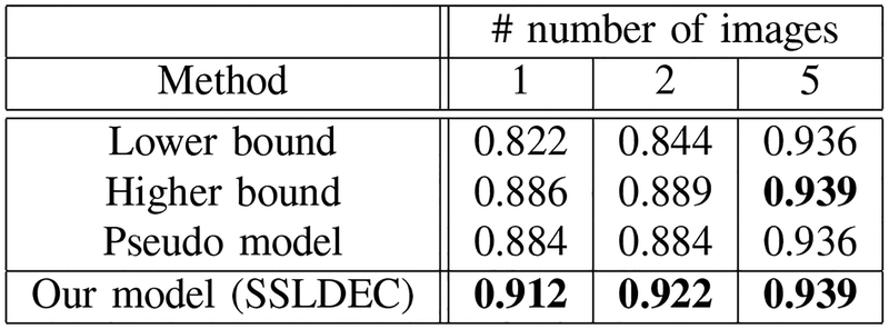
Fig. 11.
Dice scores for the cerebrospinal fluid (CSF) segmentation, compared with the lower and higher bound trained models and the pseudo-labelling semi-supervised method for experiments with 1, 2, and 5 labeled training images. For CSF segmentation our method outperformed all other models including the higher bound model which used full labeled images instead of only using one-fifth of the labels of each training image.

