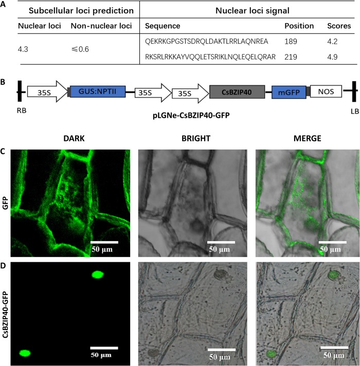
Fig 2. CsBZIP40 localizes to the nucleus.
(A) Prediction of nuclear localization with CELLO and NLS. (B) Plasmid structure for transient expression to detect the subcellular localization of CsBZIP40. LB: left border, RB: right border. Fluorescent signals of GFP in onion epidermal cells (C) were used as the control. (D) The transient expression measured by CsBZIP40-GFP fluorescence. In C and D, scale bar represents 50 μm. Each field of view was observed in dark field (DARK), bright-field (BRIGHT) and merged imaging of bright and dark fields (MERGE).

