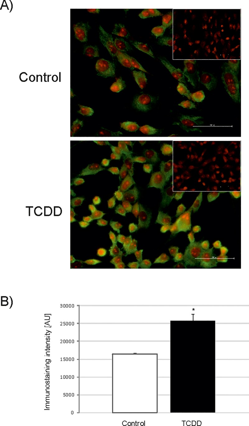
Fig 5. The abundance (mean ± SEM) of protein disulfide isomerase (PDI) in AhR-silenced porcine granulosa cells determined by immunofluorescence staining.

The immunofluorescence validation of 2D-DIGE/ mass spectrometry results was performed in untreated (Control) and TCDD-treated (TCDD) AhR-silenced granulosa cells (TR) after 12 h of incubation (n = 3 independent experiments). A) representative images of untreated (Control) cells and cells treated with TCDD (TCDD); green color depicts FITC staining of PDI and red color depicts propidium iodide staining of the nuclei. The insets represent negative controls, bar = 100 μm; brightness was enhanced for better visualization; (B) densitometric analysis of the PDI abundance; * p<0.05.
