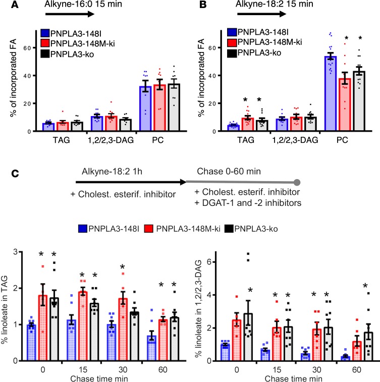
Figure 5. Partitioning of alkyne-labeled fatty acid in homozygous PNPLA3-148I, PNPLA3-148M, and PNPLA3-KO A431 cells.
(A) Cells were incubated for 15 minutes with 100 μM alkyne-palmitate and then extracted, click-reacted, and analyzed by TLC. Bars represent percentage of incorporated alkyne-palmitate in indicated lipid species ± SEM. n = 9 from 3 individual experiments. (B) Cells were incubated for 15 minutes with 100 μM alkyne-linoleate and analyzed as in A. Bars represent percentage of incorporated alkyne-linoleate in indicated lipid species ± SEM. n = 9–17 from 4–6 individual experiments; * P < 0.05 (1-way ANOVA with Dunnett’s correction). (C) Cells were incubated for a 1-hour minimum with 100 μM alkyne-linoleate in the presence of cholesterol esterification inhibitor. After labeling, cells were either collected (0 minutes chase) or further incubated in lipoprotein-deficient medium supplemented with cholesterol esterification and DGAT inhibitors for 15, 30, or 60 minutes; they were then analyzed as in A. Bars represent percentage of incorporated alkyne-linoleate in indicated lipid species, normalized to PNPLA3-148I cells at 0 minutes chase ± SEM. n = 5–8 from 3–4 individual experiments; *P < 0.05 (1-way ANOVA with Dunnett’s correction).

