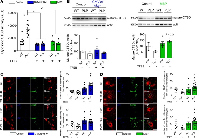
Figure 8. TFEB overexpression enhances autophagy-lysosomal pathway function in the brain of PLP mice.
(A) Quantification of Cathepsin D (CTSD) activity in cytosolic lysosomal-free fraction from ipsilateral SN of control, CMVie/hSyn-mTFEB–injected, and MBP-mTFEB–injected WT and PLP mice. (B) CTSD immunoblot levels from ipsilateral SN of control, CMVie/hSyn-mTFEB–injected, and MBP-mTFEB- injected WT and PLP mice. n = 5 per group. Lanes were run on the same gel but were noncontiguous. (C) Confocal images (left) and quantification (right) using either TH or HA tag, Lamp-2, and LC3 antibodies in the ipsilateral SN of mice injected or not with the CMVie/hSyn-mTFEB-HA. The quantification represents the number of LC3- or Lamp-2–positive puncta into neuronal cells. Scale bar: 100 μm. n = 9–13 per group. (D) Confocal images (left) and quantification (right) using either Olig2 or Flag tag, Lamp-2, and LC3 antibodies in the ipsilateral SN of mice injected or not with the MBP-mTFEB-Flag. The quantification represents the number of LC3- or Lamp-2–positive puncta into oligodendrocytes. Scale bar: 50 μm. n = 7–30 per group. White bars, control; blue bars, CMVie/hSyn-mTFEB-HA; green bars, MBP-mTFEB-3×Flag. Data represent mean ± SEM. Comparisons were made using 1-way ANOVA and Tukey’s correction for multiple comparisons. *P < 0.05 compared with control WT animals. #P < 0.05 compared with control PLP animals.

