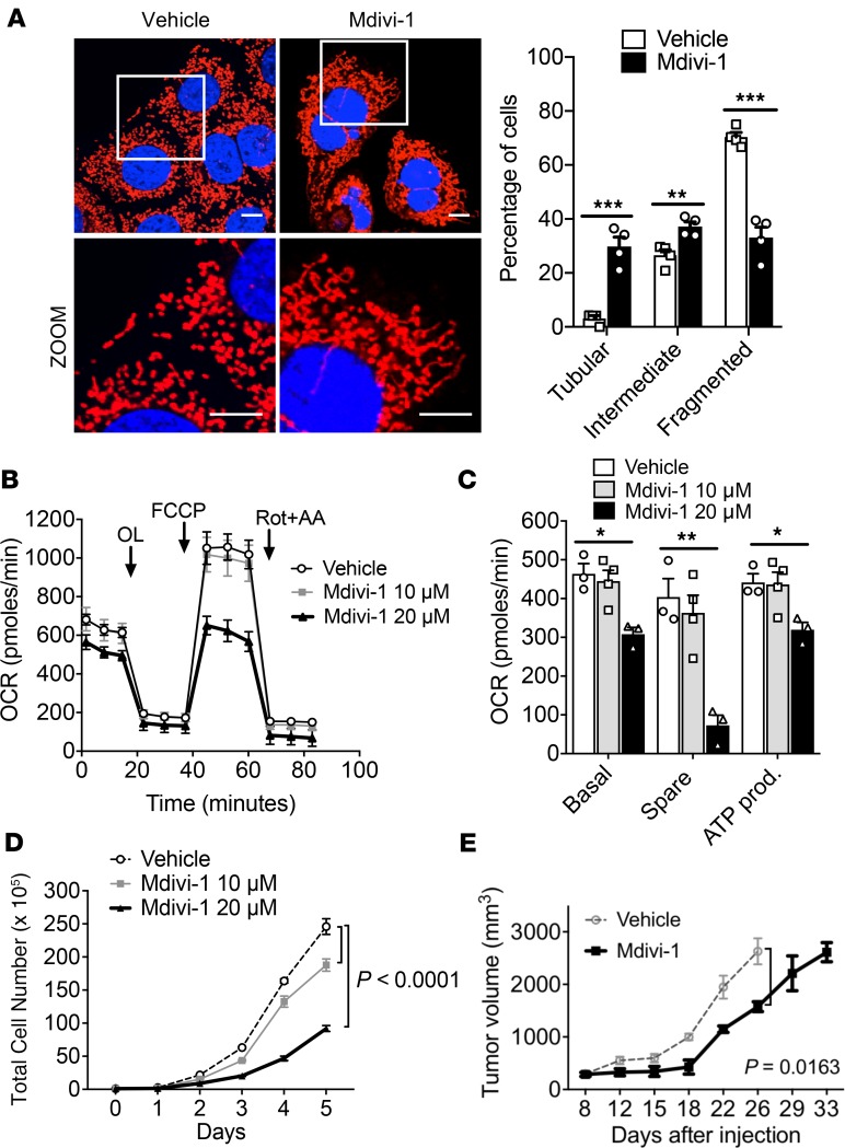
Figure 2. Suppression of mitochondrial OXPHOS by pharmacologic inhibition of mitochondrial fission improves survival.
(A) Confocal microscopy image (original magnification, ×60) of mitochondrial morphology of KPC cells treated with Mdivi-1 quantified; n = 100–200 cells. Scale bar: 10 μm. Red fluorescence, mitochondria; blue fluorescence; DAPI-labeled nucleus. ***P = 0.0006 for tubular, **P = 0.007 for intermediate, ***P = 0.0003 for fragmented by unpaired t test. (B) Mdivi-1 decreases OCR, quantified in C, and reduces cell proliferation in a dose-dependent manner (D). *P < 0.05; **P < 0.01 by unpaired t test (C); statistical analysis by 1-way ANOVA (D). (E) In vivo tumor suppression of pancreatic tumors treated with vehicle or 10 mg/kg Mdivi-1 significantly slowed tumor growth; n = 10 per cohort. Statistical analysis by 2-way ANOVA. Data are presented as mean ± SEM.

