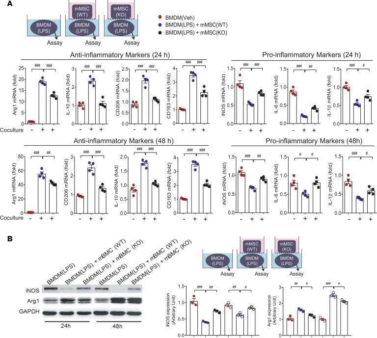
Figure 2. Deficiency of ANGPTL4 in MSCs fails to suppress inflammatory activation of macrophages.
(A) Mouse bone marrow–derived MSCs (mMSCs) were isolated from wild-type and ANGPTL4-knockout mice. In coculture settings, inflammation-related markers were assessed in LPS-stimulated mouse bone marrow–derived macrophages (BMDMs) with or without coculture with mMSCs at 24 hours and 48 hours. Wild-type mMSCs significantly suppressed proinflammatory markers, while knockout mMSCs did not. Antiinflammatory markers in macrophages were remarkably induced only by wild-type mMSCs but not by knockout mMSCs. n = 4. (B) In the same setting as A, protein levels of iNOS and Arg1 were measured by Western blot. Intensity quantification is representative of mean ± SEM. n = 3. Data are represented as mean ± SEM. #P < 0.05; ##P < 0.01; ###P < 0.001 (by Student’s t test or 1-way ANOVA with Bonferroni’s multiple-comparisons test).

