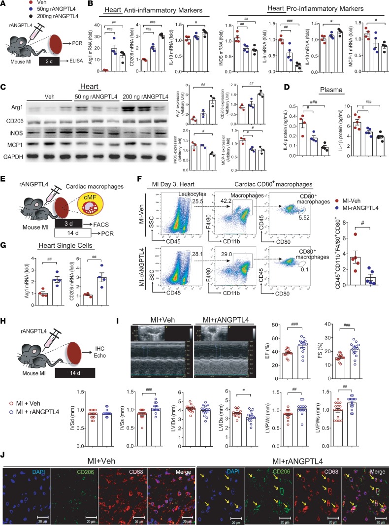
Figure 8. Cardiac inflammation and function are improved by ANGPTL4 treatment in a mouse MI model.
(A) Recombinant ANGPTL4 protein, 200 ng, was injected into the mouse MI model. Heart tissues and blood were collected after 2 days. The expression levels of inflammatory markers in the heart tissue were assessed by real-time PCR (B, n = 4) and Western blot (C, n = 3). Intensity quantification is representative of mean ± SEM. (D) Circulating IL-6 (n = 4) and IL-1β (n = 5) were reduced in ANGPTL4 protein–injected mice. (E) Experimental procedure for analyses of cardiac macrophages at 3 days or 14 days after MI. (F) The CD45+CD11b+F4/80+CD80+ cells were isolated and quantified from heart tissues at 3 days after injections of ANGPTL4 protein. n = 5. (G) The mRNA levels of Arg1 and Cd206 were assessed in the single cells isolated from heart tissues at 14 days after injections of ANGPTL4 protein or Veh. n = 4. (H) MI was induced by coronary artery ligation, then 200 ng ANGPTL4 protein was injected i.p. Heart tissues were collected after 14 days. (I) Representative echocardiogram and echocardiographic indexes expressed as graphs. n = 16 for Veh group; n = 14 for ANGPTL4 group. (J) Representative confocal images of CD68+ macrophages costained with CD206 in the infarcted myocardium. n = 8 for each group. EF, ejection fraction; FS, fractional shortening; IVSd, intraventricular septal width in diastole; IVSs, intraventricular septal width in systole; LVIDd, left ventricular internal dimension in diastole; LVIDs, left ventricular internal dimension in systole; LVPWd, left ventricular posterior wall thickness in diastole; LVPWs, left ventricular posterior wall thickness in systole. Data are represented as mean ± SEM. #P < 0.05; ##P < 0.01; ###P < 0.001 (by Student’s t test or 1-way ANOVA with Bonferroni’s multiple-comparisons test).

