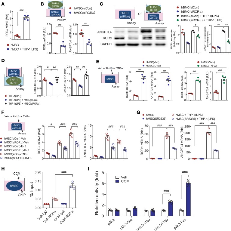
Figure 9. RORα positively regulates ANGPTL4 induction in MSCs.
(A) Rora mRNA expression was assessed in hMSCs with or without coculture with THP-1 macrophages. n = 4. hMSCs were transfected with control siRNA or RORα siRNA before coculture with THP-1 macrophages, and expression levels of RORα and ANGPTL4 mRNA (B, n = 4) and protein (C, n = 3) were measured in hMSCs. (D) Expression levels of CXCL10 and CXCL11 in macrophages were assayed after coculture with hMSCs transfected with control siRNA or ANGPTL4 siRNA. n = 4. (E) The mRNA levels of Rora and Angptl4 were assessed in hMSCs treated with IL-1β or TNF-α. n = 4. (F) The mRNA levels of Rora and Angptl4 were assessed in siRORα-transfected hMSCs treated with IL-1β or TNF-α. n = 4. (G) hMSCs were treated with SR1001, an inverse agonist of RORα and RORγ, or SR3335, a selective inverse agonist of RORα, with or without coculture with THP-1 macrophages. Inductions of RORα and ANGPTL4 were assessed by real-time PCR. n = 4. (H) hMSCs were stimulated with Veh or CCM, and binding of RORα to ANGPTL4 promoter was determined by ChIP assay. n = 3. (I) Deletion analysis of the ANGPTL4 promoter activity. HEK293T cells were transfected with the ANGPTL4-LUC reporter constructs and stimulated with Veh or CCM to analyze promoter activities. n = 4. Data are represented as mean ± SEM. #P < 0.05; ##P < 0.01; ###P < 0.001 (by Student’s t test or 1-way ANOVA with Bonferroni’s multiple-comparisons test).

