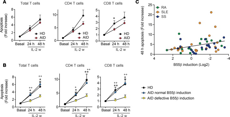
Figure 2. CWID is impaired in T cells that fail to upregulate B55β.
(A) Apoptosis was quantified by flow cytometry in activated T lymphoblasts from patients with AID and HDs, before (basal) and after IL-2 withdrawal (24 and 48 hours). Results are expressed as mean + SEM of annexin V+ DAPI− cells. *P < 0.05, two-tailed t test (HD n = 19; AID n = 59). (B) Apoptosis was compared in T cell subsets from HDs (n = 19) and from patients with AID with normal (n = 30) or defective (n = 25) B55β upregulation at 24 hours of IL-2 w. +P < 0.05 vs. HDs; ++P < 0.01 vs. HDs; ***P < 0.001 vs. AID with normal B55β induction; 2-way ANOVA with Bonferroni’s posttest. (C) Correlation between B55β induction at 24 hours (fold change over basal) and T cell apoptosis at 48 hours (fold change over basal). Spearman’s r = 0.39, and P = 0.004.

